Cataract Procedure
RECOVERY AFTER CATARACT SURGERY
At Seibel Vision Surgery, cataract surgery is a state-of-the-art procedure, using a microincisional procedure, called phacoemulsification.
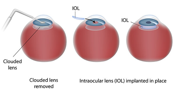
Phacoemulsification uses ultrasound to dissolve the cataract and then a tiny foldable lens is implanted in its place.
Dr. Seibel operates while looking through a highly specialized microscope; he uses the principles of his textbook Phacodynamics to enable a customized procedure for each eye of each individual patient to help insure the gentlest, safest, and most effective operation.

STEP BY STEP:
The eyelids are gently opened using the Seibel 3-D Eyelid Speculum. A very small, beveled incision, about 3 millimeters in size – less than 1/8″ wide, is made at the edge of the cornea, the transparent covering of the front of the eye. Because of the careful construction of this incision, and its small size, the incision is generally self-sealing. This translates to a “no-stitch” operation. The eye is stabilized during this step with the Seibel Gravity Fixation Ring instrument.
- Dr. Seibel then creates an opening in the capsule, which is a micro-thin membrane surrounding the cataract; he uses the Seibel Rhexis Ruler instrument to create this Continuous Curvilinear Capsulorhexis. This procedure, called capsulorhexis, requires extraordinary precision since the capsule is only about four-thousandths of a millimeter thick. This membrane is actually thinner than a red blood cell and Dr. Seibel must delicately remove the capsule while manipulating instruments within the anterior chamber – a space only 3 millimeters deep. Some studies suggest that an expertly created manual capsulorhexis can be even stronger than one created by a femtosecond laser.
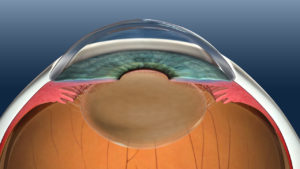
- Through the tiny incision, a microsurgical, ultrasonic, oscillating probe is inserted, which gently dissolves the cloudy lens, using high frequency sound waves. The need for ultrasound energy is minimized by the use of Dr Seibel’s instruments, the Seibel Nucleus Chopper and Safety Quick Chopper, which allow gentle disassembly of the cataract prior to ultrasound application.
- Simultaneously, this same instrument suctions out the fragmented pieces using ultrasound power. This process is called phacoemulsification, sometimes referred to as phaco. Dr Seibel applies the principles of his textbook Phacodynamics to allow the gentlest, safest, and most effective surgery that is customized to each individual patient. Furthermore, he is among the very few surgeons to utilize advanced Dual Linear Pedal control for the utmost finesse in applying machine energy for the operation.
Once the denser central nucleus of the cataract has been removed, the softer peripheral cortex of the cataract is removed using an irrigation/aspiration handpiece. The posterior capsule, an elastic bag-like membrane that held the lens, is left in place. It will support the new lens implant, as well as to maintain separation between the front and back parts of the eye.
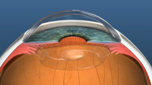
- The intraocular lens is folded and passed through the tiny incision where it is then opened (implanted) inside the capsular bag. The lens is inserted via an injector designed to help keep the incision size small – while allowing implantation of a 6 millimeter lens through a 3 millimeter (or even smaller) incision. The springy arms of the IOL, known as haptics, hold the lens implant within the capsular bag. The IOL does not generally require sutures to remain in good position.
- The lens is held in the same position as that of the natural lens (cataract) of the eye, within the capsular bag. At this stage, the cataract operation with IOL implantation is complete.
- The incision is called self-sealing because the eye’s natural internal pressure holds the incision tightly closed allowing the eye to heal without stitches. The chances of developing new astigmatism (distorted vision) after surgery are significantly decreased by eliminating stitches, which tend to pull the eye’s surface slightly out of its natural shape.
 The eyelids are gently opened using the Seibel 3-D Eyelid Speculum. A very small, beveled incision, about 3 millimeters in size – less than 1/8″ wide, is made at the edge of the cornea, the transparent covering of the front of the eye. Because of the careful construction of this incision, and its small size, the incision is generally self-sealing. This translates to a “no-stitch” operation. The eye is stabilized during this step with the Seibel Gravity Fixation Ring instrument.
 Once the denser central nucleus of the cataract has been removed, the softer peripheral cortex of the cataract is removed using an irrigation/aspiration handpiece. The posterior capsule, an elastic bag-like membrane that held the lens, is left in place. It will support the new lens implant, as well as to maintain separation between the front and back parts of the eye.